×
![Buchcover ISBN 9783642675676]()
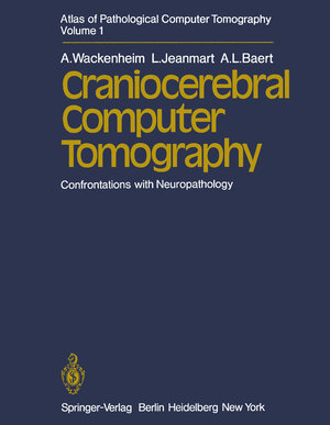
Atlas of Pathological Computer Tomography
Volume 1: Craniocerebral Computer Tomography. Confrontations with Neuropathology
von A. Wackenheim, L. Jeanmart und A.L. BaertInhaltsverzeichnis
- 1 Malformations.
- 1.1 Ventricles.
- 1.2 Hydrocephalus.
- 1.3 Bourneville Disease.
- 1.4 Aneurysms.
- 1.5 Arteriovenous Aneurysms (Angiomas).
- 2 Infections.
- 2.1 Abscesses and Empyema.
- 2.2 Abscesses and Hematoma.
- 2.3 Ventriculitis.
- 2.4 Tuberculous Meningitis.
- 3 Hematomas.
- 3.1 Intracerebral Hematomas.
- 3.1.1 Chronic Vascular Disease.
- 3.1.2 Multiple Hematoma.
- 3.1.3 Coffee Bean Hematoma.
- 3.1.4 Septate Hematoma.
- 3.1.5 Cockade-Shaped Hematoma.
- 3.1.6 “Geometric” Hematoma.
- 3.1.7 Associated Intra- and Extracerebral Hematomas.
- 3.1.8 Cortical Rupture.
- 3.1.9 Hematomas of the Posterior Fossa.
- 3.1.10 Edema Surrounding the Hematoma.
- 3.1.11 Supracallosal or Butterfly-Shaped Hematomas.
- 3.1.12 Unexplained Phenomena.
- 3.2 Intraventricular Hemorrhage.
- 3.2.1 Origin of the Hemorrhage.
- 3.2.2 Direction of the Blood Diffusion: Craniocaudal or Caudocranial?.
- 3.2.3 Intraventricular Blood Density Determination.
- 3.3 Subarachnoid Hemorrhage.
- 3.3.1 Intracerebral Hematoma with Subarachnoid Hemorrhage.
- 3.3.2 Hemorrhage in the Optochiasmatic Cistern.
- 3.3.3 Hemorrhage in the Cisterns of the Fissure of Bichat.
- 3.4 Subdural and Extradural Hematomas.
- 3.5 Hematoma and Tumor.
- 4 Ischemias.
- 4.1 Temporal Herniation.
- 4.2 “Luxury Perfusion”.
- 4.3 Postischemic Cerebral Atrophy.
- 4.4 Multiple Ischemic Lesions.
- 5 Atrophies.
- 6 Gliomas.
- 7 Metastases.
- 8 Tumor of the Pituitary Area.
- 9 Reticulosarcomas.
- 10 Ependymomas.
- 11 Pinealomas.
- 12 Meningiomas.
- References.


